During prenatal examinations, 27-year-old pregnant woman, N.P.P.A., from District 3, Ho Chi Minh City, was diagnosed with fetal heart abnormalities at 21 weeks of pregnancy, specifically progressive aortic valve stenosis. She underwent amniocentesis for genetic testing, which showed no abnormalities.
    |
 |
|
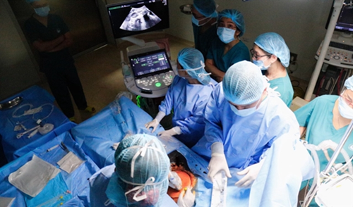
Doctors perform the fetal cardiac intervention on January 4. (Photo Courtesy of Ho Chi Minh City's Department of Health) |
By January 11, the pregnancy had progressed to 29 weeks with severe aortic valve stenosis. After consultations, fetal experts and pediatric cardiologists unanimously concluded that emergency fetal intervention was crucial in this case.
Without intervention or if delayed until after 29 weeks of gestation to dilate the aortic valve, the risk of fetal death would be extremely high.
The fetus faced the possibility of either dying in the womb, with a stillbirth rate exceeding 30%, or progressing to hypoplastic left ventricle syndrome with a 50% risk of a univentricular heart wall (requiring multiple surgeries after birth for temporary return to univentricular circulation or complete treatment with a heart transplant).
Cardiologists determined that fetal cardiovascular intervention at this juncture was appropriate. However, predicting the fetal position for cardiac catheterization presented challenges due to excess amniotic fluid and frequent fetal position changes.
The surgery started at 9am on January 12.
The surgical team faced the expected challenges, with the fetus frequently changing positions, making it difficult to insert the needle into the left ventricle and onto the aortic valve.
The intervention at Tu Du Hospital took 20 minutes to position the needle correctly, and it was then handed over to the heart valve catheterization team at Children's Hospital 1 to complete the crucial final step – aortic valvuloplasty.
Post-surgery, the pregnant woman was closely monitored in the operating room for 15 minutes, and the stabilized condition of fetal pericardial effusion was confirmed.
The successful surgery concluded at 11am on the same day, with continuous monitoring of the pregnant woman post-surgery. By 1pm, the fetal pericardial effusion was well controlled, the fetal heart rate was normal, and the mother's condition was stable.
Earlier on January 4, the same expert team from Tu Du Hospital and Children's Hospital 1 achieved a milestone by conducting Vietnam’s first fetal interventional cardiac catheterization. This involved a pregnant woman who, during her first pregnancy, was monitored in Da Nang city. Transferred to Tu Du Hospital due to the fetus's severe heart abnormalities – a birth defect without a pulmonary valve opening and right ventricular hypoplasia – the intervention successfully addressed the issues.
Subsequent examinations showed good flow through the fetal pulmonary valve, with no pericardial effusion.
Both interventions were executed with absolute precision, marking a significant advancement in technical expertise comparable to that of developed countries in the region.
Through the initial two fetal interventional cardiac catheterizations and first-hand observation of the seamless coordination by obstetric and pediatric experts at Tu Du and Children's Hospital 1, the Ho Chi Minh City Department of Health said several decisive factors contributed to the success of these interventions.
These include accuracy in ultrasound techniques by pediatric cardiologists at Children's Hospital 1, demonstrating precision in diagnosing fetal congenital heart lesions, serving as the crucial guide for accurate fetal cardiac catheterization intervention.
Experience and precision in fetal intervention performed by doctors from the two hospitals are also crucial factors.
The Ho Chi Minh City Department of Health views this success as an initial outcome in the specialized field of fetal congenital heart intervention.
Source: VNA